canis - Articles
Eye: keratoconjunctivitis sicca


Synonym(s): Dry eye
Introduction
- Ocular surface disease resulting from deficient production of aqueous phase of precorneal tear film.
- May involve decreased production of lipid or mucin portions of tear film, leading to qualitative (rather than quantitative) tear film deficiency (more difficult to diagnose).
- Cause: congenital lacrimal gland asplasia or hypoplasia; secondary to systemic metabolic disease (diabetes mellitus), drug induced (sulphonamide antibiotics, etogesic, general anesthetics, sedatives, anticholinergics); damage to local parasympathetic innervation (trauma, local inflammation, radiation therapy); auto-immune destruction of lacrimal gland; idiopathic.
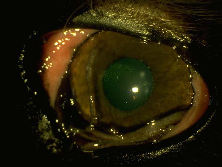
- Signs: mucoid to mucopurulent ocular discharge; conjunctival hyperemia; lackluster ocular surface; corneal edema, vascularization and pigmentation (depending on chronicity); corneal ulceration (potentially recurrent).
- Diagnosis: combination of clinical signs and low Schirmer tear test (<15 mm wetting/min) is diagnostic for quantiative deficiency of KCS. Qualitative deficiency (lipid and/or mucin layers) leads to clinical signs with normal STT, and is definitely diagnosed via tear film break-up time (uncommonly performed).
- Treatment: palliative medical therapy consisting of topical artificial tears, prophylactic antibiotics (if sedonary bacterial infection present), and/or anti-inflammatories. Treat underlying cause if possible. Depending upon underlying cause, topical cyclosporine A or oral pilocarpine to stimulate tear production. Surgical therapy (parotid duct transposition) in non-responsive cases.
- Prognosis: if congenital, unlikely to respond to medical management. May be reversible if secondary to systemic disease. If drug-induced, may be temporary/reversible or irreversible. If due to interrupted parasympathetic innervation, may be responsive to medical management. Auto-immune cases in susceptible breeds generally progressive, life-long treatment necessary. Loss of eye rare, with good owner compliance and monitoring. Print off the owner factsheet Keratoconjunctivitis sicca ('Dry eye') to give to your client.
Presenting signs
Early clinical signs- Tenacious mucoid ocular discharge, often copious, poorly responsive to topical antibiotics and/or anti-inflammatories.
- Conjunctival hyperemia.
- Blepharospasm.
- Lackluster precorneal tear film.
- Corneal edema.
- Corneal ulceration.
- Corneal vascularization and pigmentation.
Acute presentation
- Superficial non-healing corneal ulcer Cornea: spontaneous chronic corneal epithelial defects (SCCEDs).
- Deep (infected) corneal ulcer.
Age predisposition
- Middle aged to older dogs.
Breed/Species predisposition
- West Highland White Terrier West Highland White Terrier.
- American Cocker Spaniel American Cocker Spaniel.
- Lhasa Apso Lhasa Apso.
- Yorkshire Terrier Yorkshire Terrier.
- Cavalier King Charles Spaniel Cavalier King Charles Spaniel.
- Bulldog Bulldog.
- Pug Pug.
- Shih Tzu Shih Tzu.
Cost considerations
- Parotid duct transposition is expensive.
- Medical therapy is lifelong, therefore also expensive.
Pathogenesis
Etiology
- Congenital - aplasia/hypoplasia of lacrimal and/or nictitans gland. Seen in Cavalier King Charles Spaniels, Yorkshire terriers.
- Acquired:
- Immune-mediated.
- Drug-induced (sulfonamides, salicylic acid, topical atropine).
- Metabolic disease (hypothyroidism Hypothyroidism, diabetes mellitus, hyperadrenocorticism).
- Neurogenic (damage to facial VII nerve, carries motor parasympathetic fibers to lacrimal and nictitans glands)
 .
. - Radiotherapy.
- Systemic infectious disease (canine distemper Canine distemper disease.
- Trauma involving lacrimal gland (bite wounds, skull or orbital fractures).
- Idiopathic.
- Iatrogenic - surgical removal of nictitans gland.
Predisposing factors
- Breed predisposition.
Pathophysiology
- In the dog, lacrimal gland produces 70% of aqueous phase of precorneal tear film and nictitans gland produces 30% with minor contributions from modified sweat glands in lid margin (glands of Moll).
- Partial or total absence or destruction of lacrimal gland +/- nictitans gland reduced production of aqueous phase of precorneal tear film (ptf).
- Compensatory overproduction of mucous phase.
- Secondary infection resulting from loss of bacteriostatic/bacteriocidal factors in aqueous phase, eg lysozyme.
- Decreased production of outer lipid layer of tear film leads to premature evaporation of aqueous portion, while decreased production of inner mucin layer leads to poor adherence to ocular surface. These conditions lead to qualitative tear film deficiency, which has similar signs to quantitative deficiency, but may have normal STT result.
Timecourse
- Can be acute in onset or more insidious.
Diagnosis
Subscribe To View
This article is available to subscribers.
Try a free trial today or contact us for more information.
Treatment
Subscribe To View
This article is available to subscribers.
Try a free trial today or contact us for more information.
Prevention
Subscribe To View
This article is available to subscribers.
Try a free trial today or contact us for more information.
Outcomes
Subscribe To View
This article is available to subscribers.
Try a free trial today or contact us for more information.
Further Reading
Publications
Refereed papers
- Recent references from PubMed and VetMedResource.
- Sansom J, Barnett K C, Neuman W et al (1995) Treatment of keratoconjunctivitis sicca in dogs with cyclosporine ophthalmic ointment- a European clinical trial field. Vet Rec 137 (20), 504-507 PubMed.
- Stiles J, Carmicheal P, Kaswan R et al (1995) Keratectomy for corneal pigmentation in dogs with cyclosporine responsive chronic keratoconjunctivitis sicca. Vet Comp Ophthal 5 (1), 25-34 VetMedResource.
- Morgan R V & Abrams K L (1991) Topical administration of cyclosporine for treatment of keratoconjunctivitis sicca in dogs. JAVMA 199 (8), 1043-6 PubMed.
- Salisbury M A, Kaswan R L & Ward D A et al (1990) Topical application of cyclosporine in the management of keratoconjunctivitis sicca in dogs. JAAHA 26 (3), 269-74 VetMedResource.
Other sources of information
- Gelatt K N (2013)Veterinary Ophthalmology.5th edn. John Wiley & Sons.